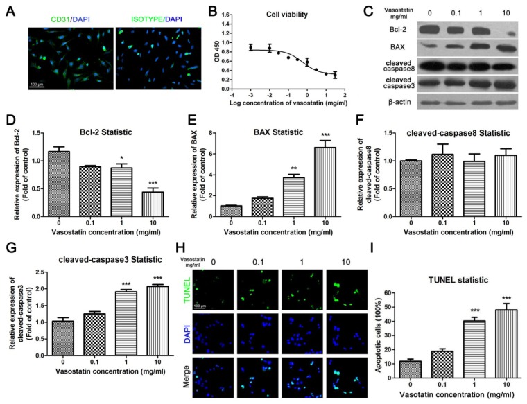
Figure 1.
Vasostatin inhibited cell viability and induced apoptosis of HUVEC under oxygen-deprivation. (A) Identification of HUVEC. Primary cultured HUVEC were stained by CD31 antibody or isotype IgG (Green), the nuclei of the cells were stained blue. Scale bar = 100 μm and referred to the two panels; (B) Cell viability assay. Cultured HUVEC were incubated with different concentrations of vasostatin under oxygen deprivation for 24 h. The absorbance at a wavelength 450 nm was measured as relative cell viability, n = 6, non-linear regression was performed to calculate IC50; (C) Determination of apoptotic markers. The expression of Bcl-2, BAX, cleaved caspase8 and cleaved caspase3 were determined by western blot analysis. β-actin was used as a housekeeping protein; (D–G) Statistical analysis of blots. All blots were performed for at least three times. * p < 0.05, ** p < 0.01, *** p < 0.001 compared to control group; (H) TUNEL staining. TUNEL-positive cells were marked by green fluorescence. The nuclei of cells were stained by DAPI. The merged figures were shown in below. Scale bar = 100 μm and refers to all panels; (I) Statistical analysis of TUNEL. The apoptotic ratio of each group was calculated as the number of TUNEL-positive cells divided by total cell number (DAPI positive); *** p < 0.001 compared to vehicle-treated group, n = 10.

