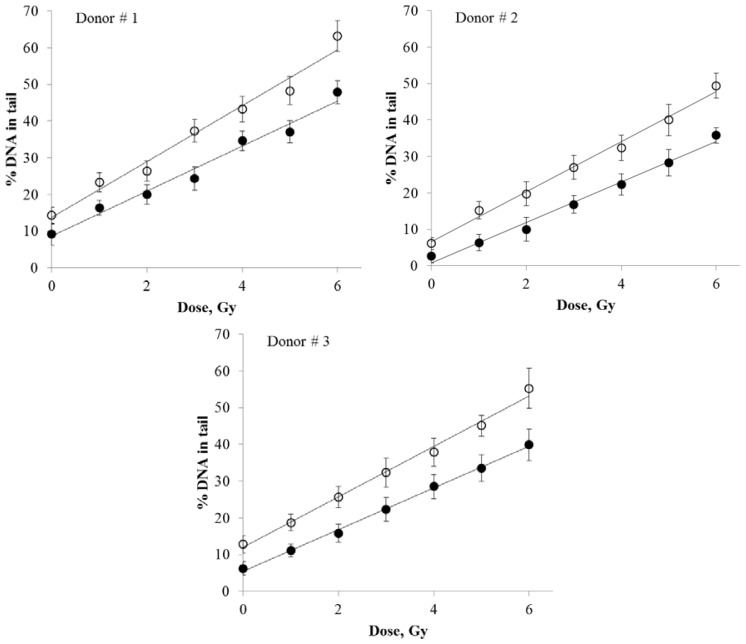
Figure 3.
The dose response curves for DNA damage in X-ray irradiated human peripheral blood lymphocytes from three donors measured by quantifying the percentage of DNA in comet tails. DNA comets prepared as described in Materials and Methods were stained with either SybrGreen I (open circles, curve 1) or Giemsa (closed circles, curve 2). The data were fitted with linear regressions. Means of three measurements (three slides per treatment) ± standard errors are shown.

