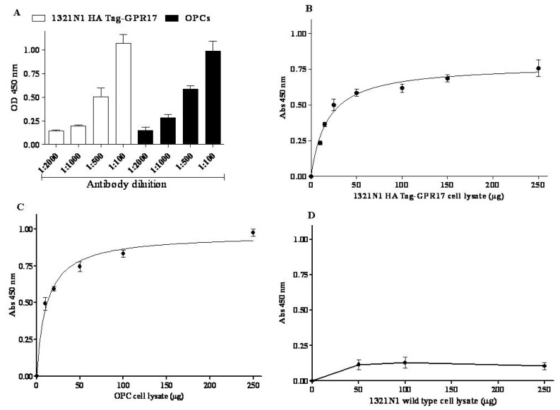
Figure 1.
(A) Cell lysates (10 μg) obtained from 1321N1 HA Tag-GPR17 cells (white bars) or primary OPCs (black bars) were captured on wells pre-coated with anti-HA Tag antibody or anti-N terminus-GPR17 antibody, respectively. Different-fold dilutions of a full-length anti-GPR17 antibody were probed in 120 min blocking time. The secondary HRP-conjugated antibody and the TMB substrate kit allowed a colorimetric quantification of signals. Blanks were obtained processing cell lysates in the absence of the primary anti-GPR17 antibody; (B,D) Different amount of cell lysates obtained from 1321N1 HA Tag-GPR17 (B), or 1321N1 wild type cells (D) were captured on wells pre-coated with anti-HA Tag antibody. After extensive washes, levels of GPR17 were quantified using a full-length anti-GPR17 antibody, and subsequently an HRP-conjugated antibody/TMB substrate kit; (C) Different amounts of cell lysates obtained from primary OPCs were captured on wells pre-coated with anti-N terminus-GPR17 antibody. After extensive washes, levels of GPR17 were quantified using a full-length anti-GPR17 antibody, and subsequently an HRP-conjugated antibody/TMB substrate kit. Blank wells were obtained in the absence of the full length anti-GPR17 antibody. Data are expressed as specific absorbance at 450 nm and represent the mean ± SEM of at least three independent experiments.

