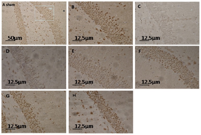
Figure 1.
Light micrograph of hippocampus sections in sham- and TBI-rats at different time-points post-injury (immunohistochemistry for S100A6). The normal structure of the hippocampus from a sham rat is depicted in (A) (10 × 10); a rectangular section of the CA2 region is enlarged in (B) (40 × 10); Normal pyramidal neurons in sham rats were interspersed with condensed dark-brown neurons. In TBI-rats, the number and density of pyramidal neurons noticeably decreased at 1 h post-injury as shown in (C) (40 × 10); and began to increase gradually from 6 h to 12 h, 24 h, 72 h, and 14 day, as shown respectively in (D–H) (40 × 10). These neurons, which are full of densely-compacted fine particles, provide strong evidence of higher cellular S100A6 immunoreactivity.

