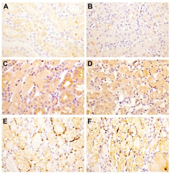
Fig. 7.

Myeloperoxidase immunohistochemistry.
Kidney sections from wild-type (A, C, E) and Cyp4f18 −/− mice (B, D, F) at 12 h (C, D) and 24 h (E, F) post-ischemia. Minimal background staining was observed in sham-operated mice (A, B). By 12 hpi there was an appreciable increase in the number of MPO positive cells. This number further increased by 24 hpi (E, F) in both the wild-type and Cyp4f18 −/− mice.
