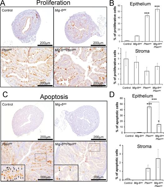
Figure 3.
The regulation of proliferation and apoptosis by Mig-6. (A) Immunohistochemical analysis of phospho-histone H3 as a proliferation marker in uteri of control, Mig-6d/d, Ptend/d, and Mig-6d/d Ptend/d mice at 2 weeks of age. (B) Quantification of phospho-histone H3 positive cells in epithelial and stromal cells (C) Immunohistochemical analysis of cleaved caspase 3 as an apoptotic cell marker. Small arrows indicate apoptotic cells. (D) Quantification of cleaved caspase 3 positive cells in epithelial and stromal cells. *, p<0.05; ***, p<0.001

