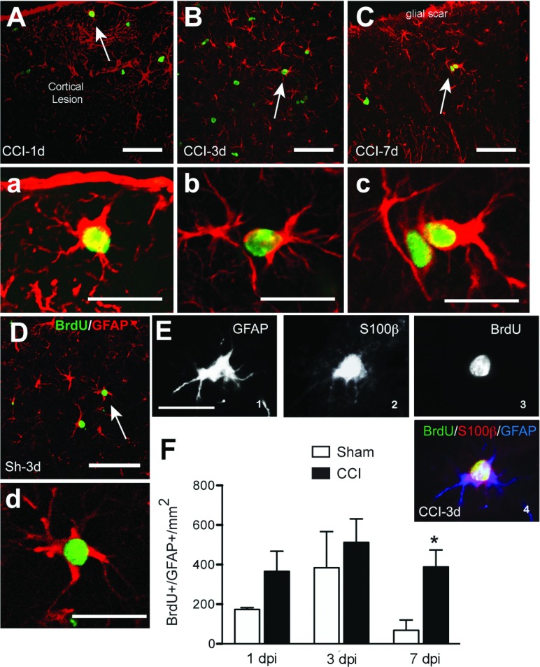
Figure 3. Astrocytic proliferation after CCI injury.
Co-localization of GFAP (red) and BrdU (green) after CCI or sham injury around the cortical lesion and in the corpus callosum at different time points. Proliferating astrocytes have the morphology of reactive astrocytes with hypertrophic cell body and processes. Arrows indicate cells shown at larger magnification in their corresponding panels. Some sections taken at 3 dpi were triple labeled for GFAP+/S100β+/BrdU+ (E1-4) confirming that dividing GFAP+ cells were astrocytes. Scale bar=50 μm (A, B, C and D) and 20 μm (a, b, c, d and E). (F) Quantification of GFAP+/BrdU+ cells at different time points after CCI or sham injury (mean±S.E.M., n=3–6). *P<0.05, **P<0.01, by two-tailed Student's t test comparing sham and CCI at each time point.

