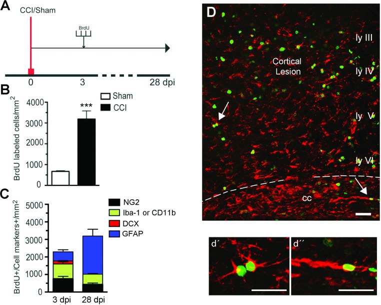
Figure 6. Fate of proliferating cells 4 weeks after CCI injury.
(A) Experimental design for BrdU injections. BrdU (100 mg/kg body weight) was injected at 3 dpi every 3 h for a total of four injections. One group of mice was killed at 3 dpi, and the second group at 28 dpi. (B) A significant increase in BrdU-labeled cells was found in the injured mice compared with sham mice at 28 dpi (***P<0.001). (C) Cell phenotype of BrdU+ cells in the ipsilateral injured cortex of mice killed at 3 or 28 dpi. At 28 dpi we observed more proliferating cells, most of which were BrdU+/GFAP+. The number of BrdU+ cells co-stained with Iba-1 or NG2 decreased, and no BrdU+/DCX+ cells were observed. (D) Confocal image showing colocalization of GFAP (red) and BrdU (green) across the cortical layers and corpus callosum at 28 dpi, and arrows point to proliferating astrocytes shown at high magnification in cortex (d’) and corpus callosum (d’’). Scale bar=50 μm (D) and 20 μm (d’ and d’). (mean±S.E.M., n=4).

