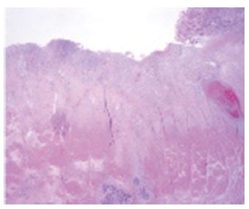
Figure 3.

Laparoscopic assisted distal gastrectomy specimen. The ulcerative lesion due to mucosal detachment after endoscopic submucosal dissection is distinguished from normal mucosa (right side). Fibrosis was observed in the submucosal layer and a hypercellular lesion that was the same as the endoscopic submucosal dissection specimen in the muscle and subserosa layers.
