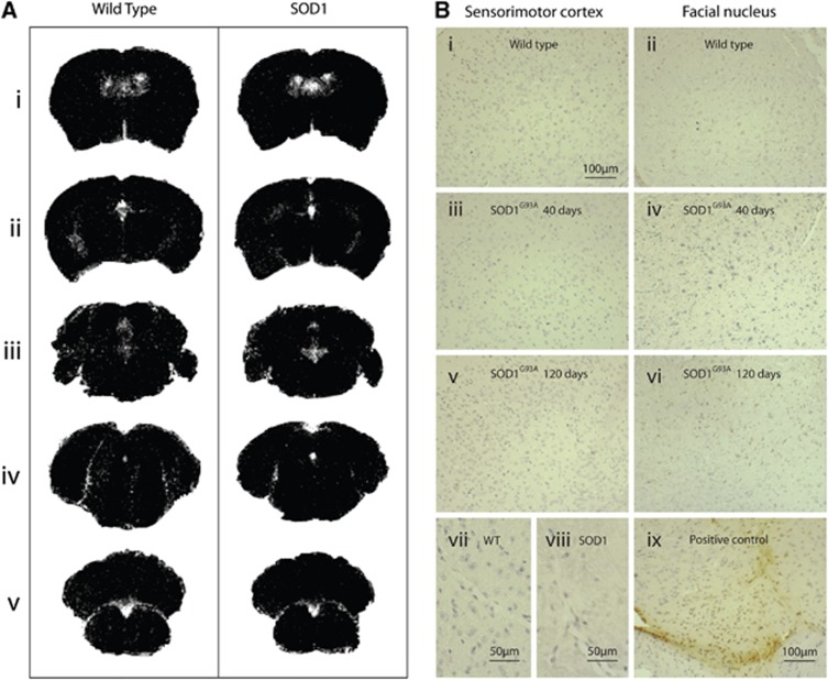
Figure 5.
No evidence of blood–brain barrier (BBB) breakdown is seen at any time point in SOD1G93A mice. Subtraction of Gadolinium T1 images (A) showed Gd-DTPA signal change in the ventricles in both SOD1G93A and wild-type (WT) mice, but no signal change is seen in the tissue, either in the sensorimotor cortex (i and ii) or in any of the brainstem motor nuclei (iii–v). No IgG staining was noted at any time point (B) in WT (i and ii), 40-day SOD1G93A (iii and iv), or 120-day SOD1G93A (v and vi) tissue. Panels i, iii, and v show images from the sensorimotor cortex; ii, iv, and vi show images from nucleus VII. Similarly even at high magnification, no staining was seen around vessels in SOD1 (viii) or WT (vii) mice. Panel ix shows a photomicrograph from positive control tissue (experimental brain trauma) as a comparison. No change was seen in the spinal cord (images not shown). N=4 in each group. SOD1, superoxide dismutase; Gd-DTPA, gadolinium diethylene triamine pentaacetic acid.

