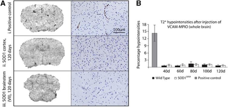
Figure 6.
Imaging of VCAM-1 expression, and the corresponding immunohistochemistry, in the brains of SOD1G93A mice and wild-type (WT) controls at 120 days, and positive interleukin-1β (IL-1β)-injected controls. Representative T2* images of the cortex (A ii, left panel) and brainstem (facial nucleus, A iii, left panel) in SOD1G93A mice at end stage show no binding of VCAM-MPIO. However, significant binding could be seen after injection of IL-1β (A i, top left), where VCAM-MPIO binding causes signal dropout. This was confirmed by VCAM immunohistochemistry (A, right panels). Quantification of T2* hypointensities (B) shows that there was a significant VCAM-MPIO binding in the positive control only, with no significant binding in SOD1G93A or WT mice at any time point. Data presented are for whole brain; analysis was also performed for the sensorimotor cortex and the brainstem in isolation, but no regional effects were found either (data not shown). Positive control N=3; SOD1G93A and WT N=4. Scale bar applies to all immunohistochemistry images. VCAM-1, vascular cell adhesion molecule 1; MPIO, microparticles of iron oxide.

