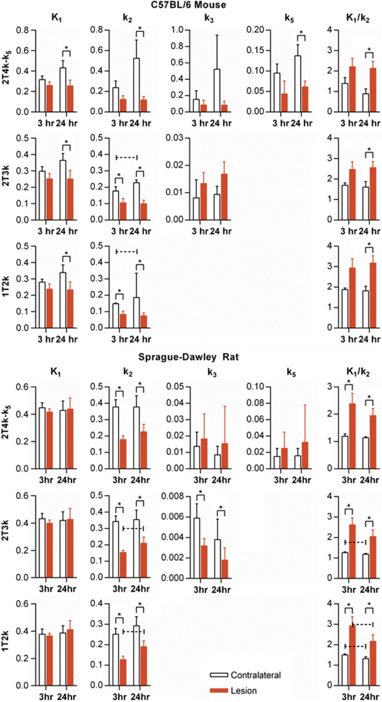
Figure 4.
[18F]fluoroacetate kinetic modeling parameters for both mouse and rat cerebral H-I for both 3 and 24 hours post insult imaging groups. 1T2k, 2T3k, and 2T4k-k5 models (shown in rows) were fit to the data. An asterisk (*) over a pair of bars indicates paired t-test of difference between contralateral versus lesion mean yielded P<0.05. A dashed, capped line over a pair of time points indicates an unpaired t-test difference between 3- and 24- hour means yielded P<0.05.

