
FIG. 1.
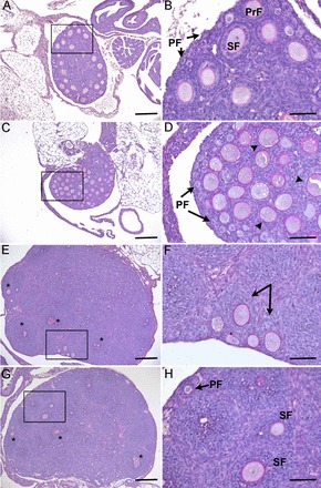
Genetic deletion of Inha and Gdf9 alters folliculogenesis at PND 12. Twelve-day-old control (A, B) ovaries contain primordial (PF) and primary (PrF) and secondary follicles (SF), while 12-day-old Gdf9−/− (C, D) ovaries block at the primary stage with a single layer of granulosa cells (arrowheads). E, F) Inha−/− ovaries are much larger at PND 12 and contain large multilayer follicles (* in E) and abnormal asymmetric secondary follicles (arrows in F). G, H) Twelve-day-old Inha−/− Gdf9−/− ovaries have a morphologically similar phenotype to Inha−/− ovaries with large multilayered follicles (* in G) that are not observed in age-matched controls or Gdf9−/− alone. Boxed regions in the left panels are shown at higher magnification in the right panels. Bars = 200 μm (A, C, E, G) and 50 μm (B, D, F, H).