
FIG. 5.
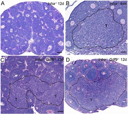
Loss of Gdf9 enhances the onset of microscopic tumor foci in Inha−/− ovaries. A) PND 12 Inha−/− ovaries do not display evidence of tumor formation until 4 wk of age. B) Black outlined region denoted as T shows tumor formation in 4-wk-old Inha−/− ovaries. C, D) Twelve-day-old Inha−/− Gdf9−/− double mutants shows regions of hyperplasia of various sizes that resemble the pretumor foci (T) observed in older Inha−/− ovaries (B). Bars = 100 μm (A, C, D) and 50 μm (B).