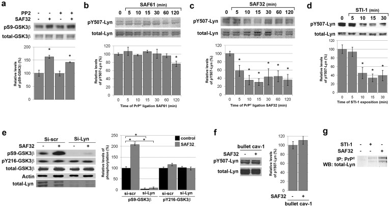
Figure 3. PrPC-mediated GSK3β phosphorylation on S9 is relayed by the Lyn kinase in 1C115-HT neuronal cells.
(a) 1C115-HT neuronal cells were pre-incubated with the Fyn inhibitor PP2 (50 pM, 1 h) prior to exposure to SAF32 anti-PrPC antibodies (10 μg/ml, 30 min), targeting native PrPC. Cell lysates were immunoblotted with antibodies targeting pS9-GSK3β and total GSK3β for normalization. (b–d) 1C115-HT neuronal cells were exposed to anti-PrPC antibodies targeting (b) a C-terminal epitope (SAF61, 10 μg/ml), (c) a N-terminal epitope (SAF32, 10 μg/ml) or (d) to a peptide corresponding to the domain of STI-1 that binds PrPC (aa 230–245) (25 μM). Cell lysates were immunoblotted with antibodies targeting pY507-Lyn and total Lyn for normalization. (e) 1C115-HT neuronal cells were transfected for 36 h with a siRNA targeted against Lyn (Si-Lyn) or a control scramble siRNA (Si-scr) prior to exposure to SAF32 anti-PrPC antibodies (30 min). Cell lysates were immunoblotted with antibodies targeting pS9-GSK3β, pY216-GSK3β, total GSK3β. Total Lyn was used to check knockdown and actin was used as loading control. (f) 1C115-HT neuronal cells were submitted to caveolin-1 immunosequestration prior to exposure to SAF32 antibodies (10 μg/ml, 15 min). Cell lysates were immunoblotted with antibodies against pY507-Lyn and total Lyn for normalization. (g) 1C115-HT neuronal cells were exposed to the STI-1 peptide (25 μM) or SAF32 antibodies (10 μg/ml) for 30 min. Cell lysates were immunoprecipitated with SAF61 anti-PrPC antibodies and immunoblotted with antibodies against Lyn. Gels have been cropped for clarity and conciseness purposes; original images corresponding to (b–c) are shown in Supplemental Figure 5. Data are expressed as means ± S.E.M of n = 4 to 6 independent analyses. *P < 0.05 vs. control, Kolmogorov-Smirnov test.

