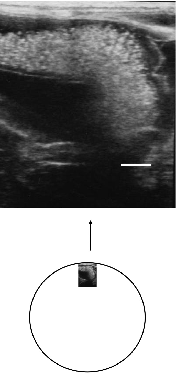
Fig. 3.

Ultrasonography performed on day 3 demonstrates a large hyperechoic mass, suggesting hematoma. As shown in a schema of whole part of hematoma as large as volley ball beside the right pregnant uterine horn. A hyperechoic area of surface part of hematoma, suggesting a clotted blood of hematoma. The inner part of hematoma indicates unclotted hematoma. Bar indicates 1 cm.
