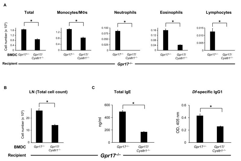
FIGURE 7. GPR17-deficient Mice Sensitized with Df-pulsed GPR17-deficient BMDCs.
(A) Inflammatory cell count in BAL fluid. BMDCs from Gpr17−/− and Gpr17/Cysltr1−/− mice were pulsed with Df at 100 μg/ml for 24 h, and 104 cells were administered intranasally for sensitization of Gpr17−/− recipients. Recipients were challenged with 1 μg of Df intranasally at days 10 and 14 and killed at day 16 for analysis. Total and differential cell counts for BAL fluid monocytes/macrophages (MΦs), neutrophils, eosinophils, and lymphocytes are shown. (B) Parabronchial LN cell numbers. Parabronchial LN cells were harvested and counted, and total cell numbers are shown. (C) Levels of total IgE and Df-specific IgG1 in serum. Total IgE and Df-specific IgG1 in sera of Df-challenged mice were measured by ELISA. Values are the mean ± SEM (n = 10) combined from 2 independent experiments. *P < 0.01.

