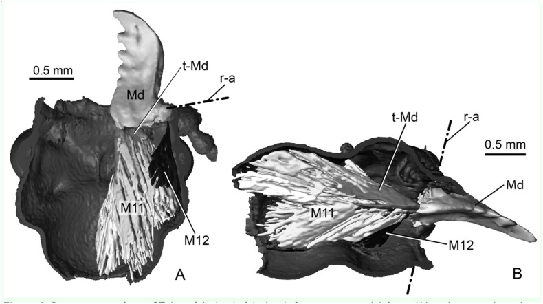
Figure 3.
Reconstructions from µCT data of the head of the beetle Priacma serrata with left mandible and associated muscles. (A) Ventral view of dorsal half of head. (B) Right-lateral view of left half of head. M11: adductor muscle; M12: abductor muscles; Md: mandible; r-a: rotation axis of mandible; t-Md: tendon of M11. High quality figures are available online.

