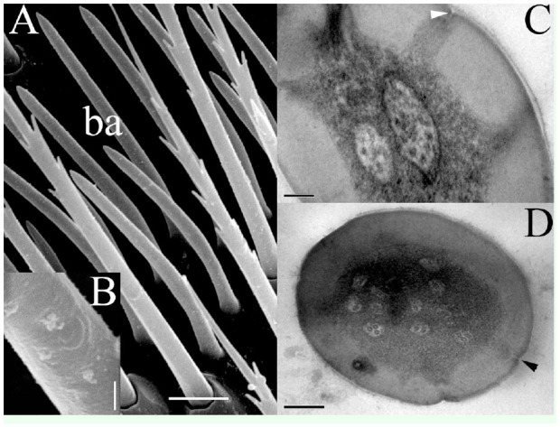
Figure 3.

(A) Sensillum basiconicum (ba) in a sensory band. (B) shows the slightly grooved surface of this type of sensilla. (C) In the cross-section, the thick wall is interrupted by spaced pores (arrowhead), which communicate outwardly using three dendrites in the lumen. (D) Several dendritic segments and pores (arrowhead) formed in the upper part of the basiconicum. Scale bars: A = 5 µm; B = 0.5 µm; C = 0.2 µm; D = 0.1 µm. High quality figures are available online.
