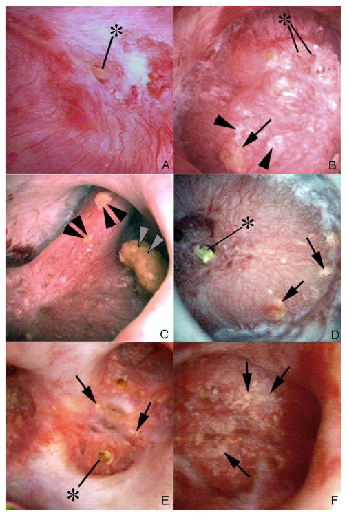
Figure 1. Endoscopic views of renal papilla from HASF.
Panel a shows a renal papilla from patient 1 that appears normal except for a protruding plug (asterisk) from a dilated opening of a duct of Bellini. No regions of plaque are noted on the surface of this papilla. In contrast, panel b shows a papilla from patient 6 with large sites of Randall’s plaque. Several protruding plugs (asterisk) are noted among numerous sites of Randall’s plaque (arrowheads) and an occasional site of yellow plaque (arrow). Panel c (patient 6) shows three separate attached stones (double arrowheads). The larger stone was determined to be attached to a region of RP and the stone composition was apatite. Panels d–f (panel d, patient 7; panel e, patient 5; panel f, patient 3) show the papillary surface of three additional CaP patients that possess regions of yellow plaque (arrows) and little to no Randall’s plaque. The two patients 5, 3 (panels e and f) showed papillary retraction and flattening as well as heavy amounts of yellow plaque (arrows). No sites of Randall’s plaque were noted in patients 3 and 5.

