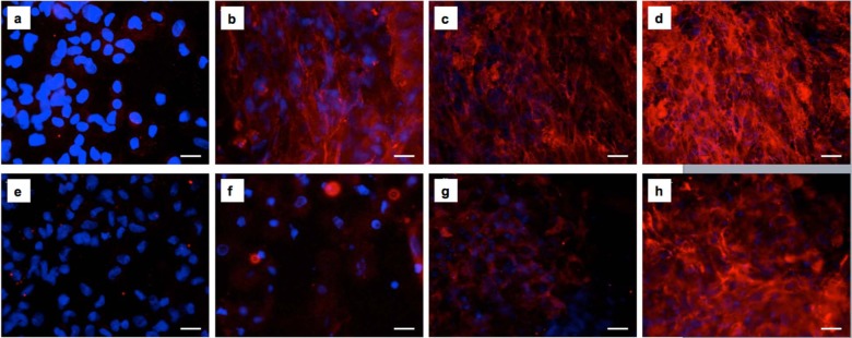
Figure 2.
Fluorescence micrographs of collagen I secretion by HDFa cells 1, 3, 7 and 14 days post culture within Fmoc-FF/RGD and Fmoc-FF/RGE hydrogels. Collagen I was deposited and organized into a dense fibrous network in the Fmoc-FF/RGD hydrogel through the culture term (a) 1, (b) 3, (c) 7 and (d) 14 days post culture. Collagen I was much less expressed in the Fmoc-RGE gel with structureless staining observed (e) 1, (f) 3, (g) 7 and (h) 14 days post culture. Blue: cell nuclei; Red: collagen I. Scale bars: 25 µm.

