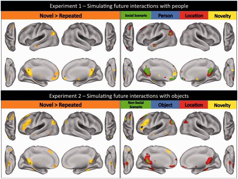
Fig. 2.
Results for Experiments 1 and 2. Top left panel: Regions of the brain demonstrating repetition-related reductions in neural activity for simulated future social events in Experiment 1 [i.e. greater activity when participants simulated future social events for the first time (i.e. novel trials) relative to a third time (i.e. repeat trials); whole-brain corrected (FWE) at P < 0.05; prominent activations include (a) medial prefrontal; (b) posterior cingulate; (c) left angular; and (d) left middle temporal cortices]. Top right panel: Regions involved in constructing simulated representations of future social events that are preferentially associated with processing details about: (1) social scenarios (prominent activations include medial prefrontal, posterior cingulate, left temporal–parietal and left middle temporal cortices); (2) people (prominent activations include medial prefrontal cortex); (3) locations (prominent activations include posterior cingulate cortex and left angular gyrus); and (4) novelty (prominent activations include anterior cingulate cortex and left hippocampus) [all activations whole-brain corrected (FWE) at P < 0.05]. Bottom left panel: Regions of the brain demonstrating repetition-related reductions in neural activity for simulated future non-social events in Experiment 2 [i.e. greater activity when participants simulated future non-social interactions for the first time (i.e. novel trials) relative to a third time (i.e. repeat trials); whole-brain corrected (FWE) at P < 0.05; prominent activations include (a) medial prefrontal; (b) retrosplenial; (c) bilateral angular; and (d) bilateral parahippocampal cortices]. Bottom right panel: Regions involved in constructing simulated representations of future non-social events that are preferentially associated with processing details about: (1) non-social scenarios [prominent activations include retrosplenial cortex and left angular gyrus (extending to occipital cortex)]; (2) objects (prominent activations include left inferior prefrontal and left premotor cortices); (3) locations (prominent activations include retrosplenial, bilateral parahippocampal and right angular cortices); and (4) novelty (prominent activations include left lateral prefrontal cortex) [all activations whole-brain corrected (FWE) at P < 0.05].

