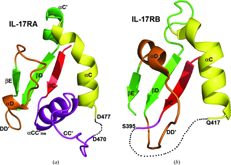
Figure 3.
IL-17RA and IL-17RB SEFIR domains adopt different topologies. Depicted are partial structures of the IL-17RA (left) and IL-17RB (right) SEFIR domains. For clarity, parts of the SEFIR structures are omitted. The black dashed lines represent disordered loops that are not observed in the structures. Notice that strand βC (red) and helix αC (yellow) in IL-17RA SEFIR are connected by a long insertion containing αCC′ins and the CC′ loop (purple) without forming a knot. In contrast, the corresponding fragment in IL-17RB SEFIR is largely disordered and connects strand βC (red) and helix αC (yellow) in a knot topology.

