Fig. 5.

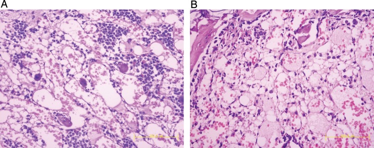
BM histological analyses of irradiated mice on Day 7 in WT mice (A) and SRC-3−/- mice (B) exposed to a TBI of 6.0 Gy (n =3) (stained with hematoxylin and eosin, ×200).

BM histological analyses of irradiated mice on Day 7 in WT mice (A) and SRC-3−/- mice (B) exposed to a TBI of 6.0 Gy (n =3) (stained with hematoxylin and eosin, ×200).