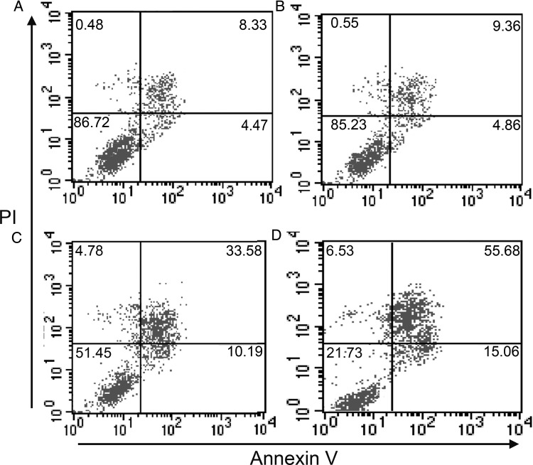
Fig. 6.
Apoptosis of BM nucleated cells in SRC-3−/- and WT mice after 6 Gy irradiation and control (no irradiation). (A) Cells collected from WT mice exposed to no irradiation. (B) Cells collected from SRC-3−/- mice exposed to no irradiation. (C) Cells collected from WT mice after irradiation. (D) Cells collected from SRC-3−/- mice after irradiation. The percentages of early apoptotic cells (annexin-high/PI-low) and apoptotic/dead cells (annexin-high/PI-high) are shown for each cell population by flow cytometry.

