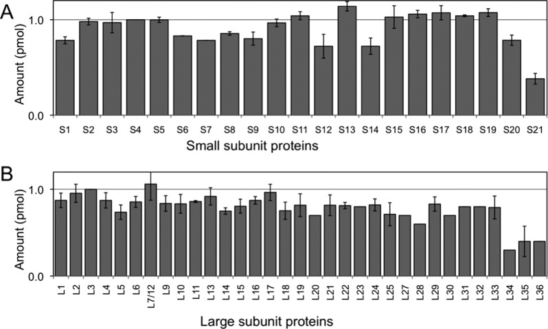
Figure 6.
Quantification of 30S r-proteins (A) and 50S r-proteins (B) using ion mobility separation and multiple protease digestion as improvements to the LC–MSE approach. A uniform distribution of proteins is observed for the majority of r-proteins quantified, which is in agreement with their known stoichiometry within the complex. Ribosomal proteins S21, L34, L35, and L36 are not quantified because they generate too few peptides, even using multiple enzymatic digestion and ion mobility separation. Amounts of small subunit and large subunit proteins are normalized to r-protein S4 and r-protein L3, respectively. Error bars represent the standard deviation of three replicate measurements.

