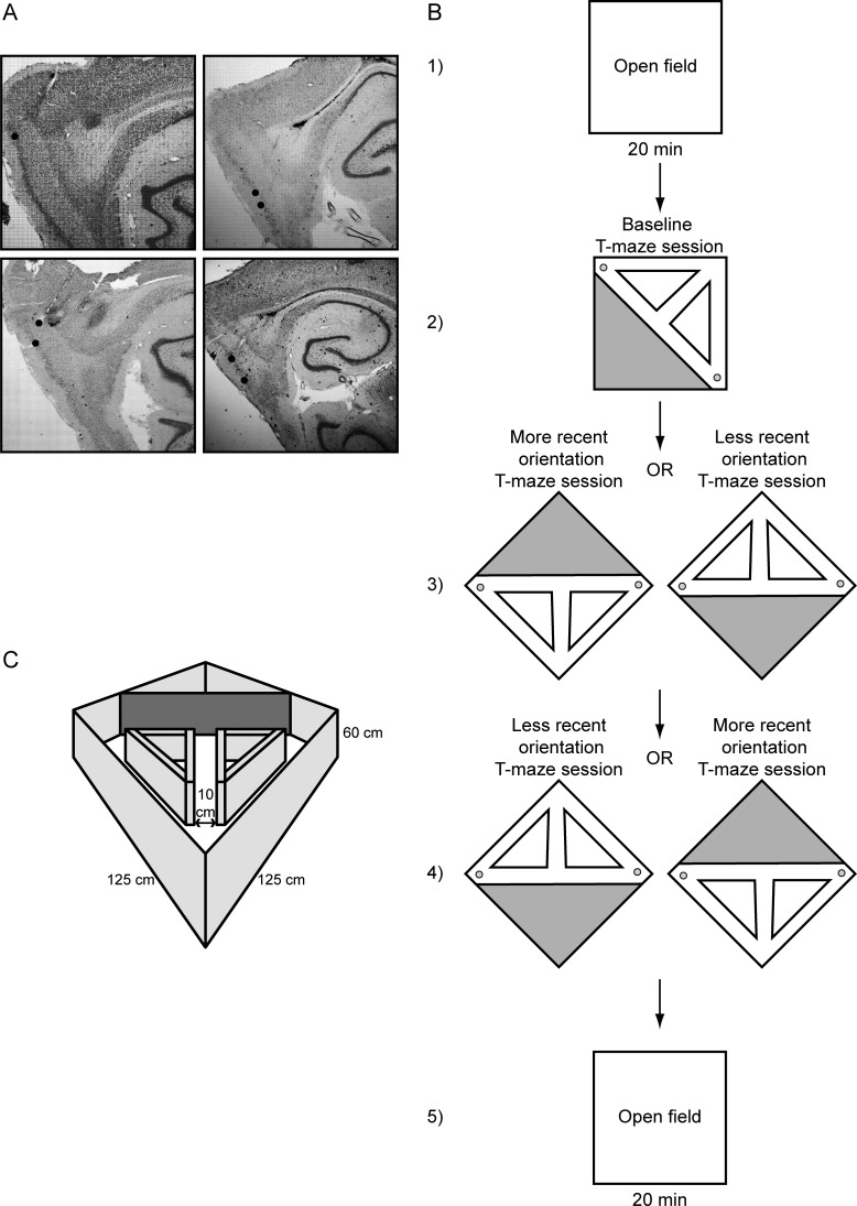
Figure 1.
(A) Nissl-stained sagittal sections showing tetrode tracks going through layers II–V of the mEC in 4 different rats. (B) The testing protocol shows an initial 20-min open-field session followed by 3 subsequent T-maze sessions and a final 20-min open-field session. (C) The testing environment showing the square open field with prisms and blocking wall generating the T-maze.

