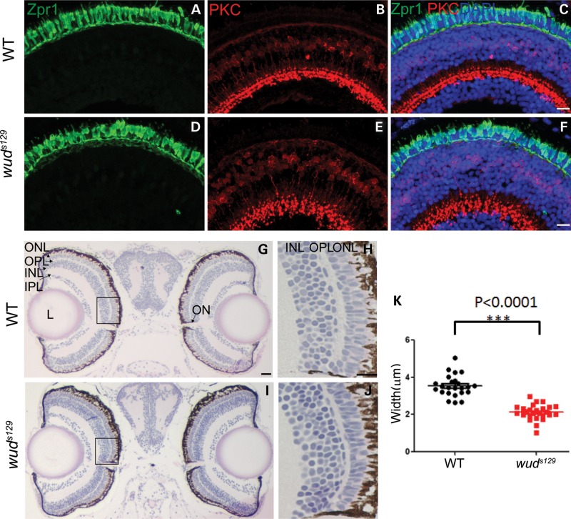
Figure 1.
wud mutants have a thinner outer plexiform layer. (A–F) IHC revealed relatively normal retinal morphology. Larvae at 7 dpf were stained with zpr1 (A and D), which labels double cones, and PKC (B and E), which labels bipolar cells. Nuclei were counterstained with DAPI in the merged images (C and F). (G–K) wud mutants have a thinner outer plexiform (OPL) layer. Shown here are H&E stained semi-thin plastic sections of wild-type (G) and wud (I) larvae at 7 dpf. Magnified views of the boxed areas in G and I are shown in H and J, respectively. Measurements (n = 24 per group) revealed a significantly thinner OPL (K, P< 0.0001). ONL, outer nuclear layer; OPL, outer plexiform layer; INL, inner nuclear layer; IPL, inner plexiform layer; L, lens; ON, optic nerve. Scale bars for C, F and H is 10 µm and G is 20 µm.

