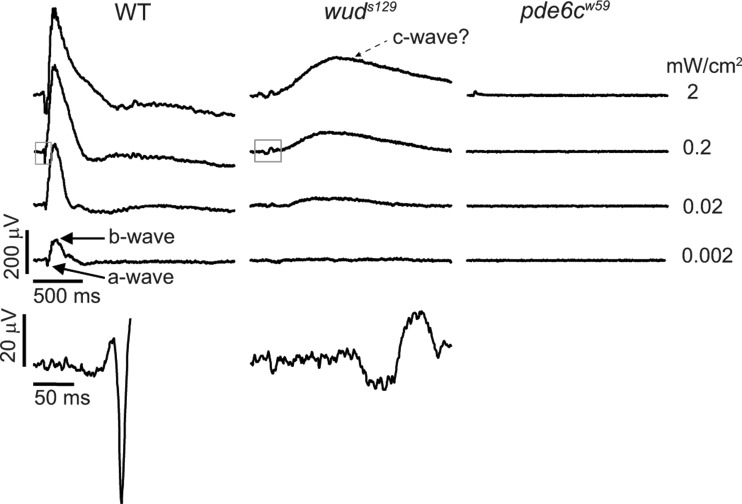
Figure 2.
wud mutants display abnormal ERGs. Wild-type larvae showed stereotypical ERGs across 4 log units of illumination (flash intensities are shown on the right). The a- and b-wave components of the ERG are indicated in the dimmest trace (bottom left). wud mutants showed defective ERGs with significantly reduced sensitivity. These waveforms comprise a small a-wave followed by a delayed and reduced b-wave and a slow, large-positive component that may represent a c-wave indicated on the top wud trace. No ERG responses were recorded from the pde6c mutants, although a miniscule flash artifact can be seen on the brightest trace (top, right trace). Close-ups of the boxed regions from the second wild-type and wud traces are shown on the bottom to illustrate the magnitude of the a-wave and the delay of the b-wave.

