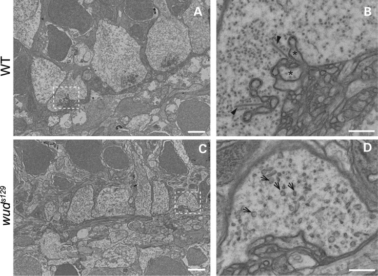
Figure 5.
Synaptic ribbons are absent in wud mutants. (A–D) Transmission electron microscopy of adult wild-type and wud mutants. In wild type (WT), the cone pedicles showed dendritic invaginations and multiple docked synaptic ribbons (A and B). In (B) horizontal cells (asterisks) and synaptic ribbons (arrowheads) are indicated. In contrast, the wud mutants had abnormal cone pedicles that lacked dendritic invaginations and synaptic ribbons (C and D). srPB were observed in the wud mutants (arrows in D). Scale bar: (A) and (C), 2 µm; (B) and (D), 500 nm.

