Figure 7.
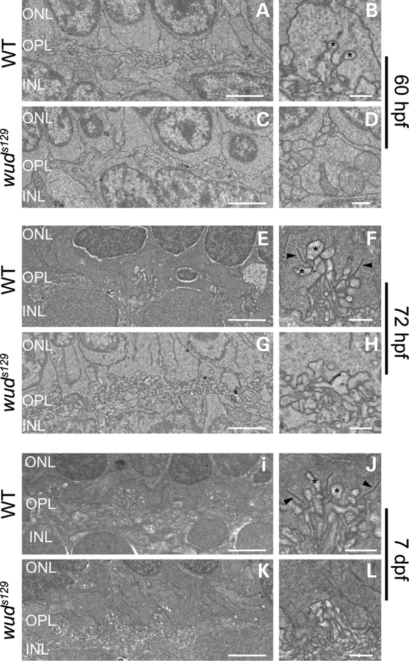
Cone synaptic ribbon formation is defective in wud mutants. Transmission electron microscopy of wild-type and wud mutant larvae during synaptic ribbon development. (A–D) At 60 hpf, no synaptic ribbons were observed in wild-type or wud mutants. Wild-type embryos showed the early stages of dendritic invagination into the cone pedicles (asterisks) (B), which was absent in the cone pedicles of wud mutants (D). At 72 hpf, wild-type embryos showed normal cone pedicles containing synaptic ribbons (E and F). wud mutants had abnormal cone pedicles and lacked synaptic ribbons (G and H), indicating a defect in the initial formation of synaptic ribbons. At 7 dpf, the results were similar to 72 hpf, where the wild-type larvae showed normal cone pedicles and synaptic ribbons (I and J) and wud mutants lacked synaptic ribbons (K and L). Arrowheads in (F) and (J) indicate synaptic ribbons. hpf, hours postfertilization; dpf, days postfertilization. Scale bar: (A), (C), (E), (G), (I) and (K), 2 µm; (B), (D), (F), (H), (J) and (L), 500 nm.

