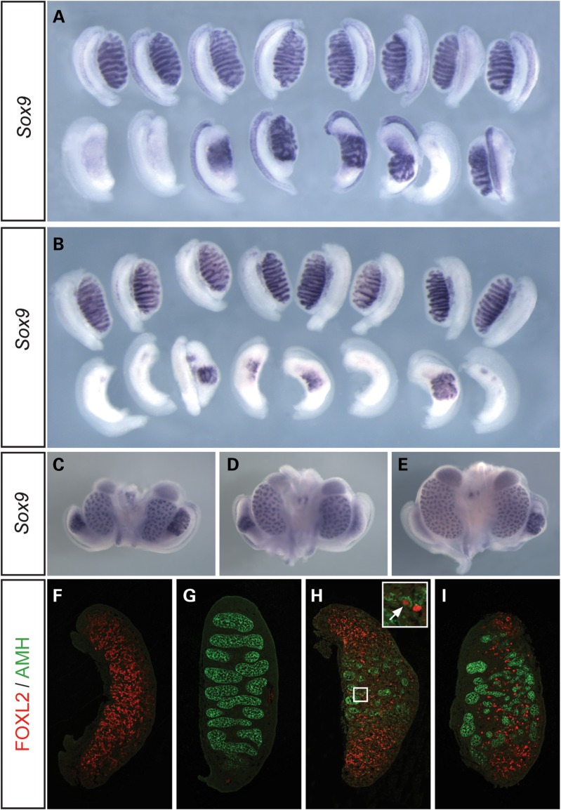
Figure 1.
Abnormalities of testis development in XY Map3k4tm1Flv/+ and XY Thp/+ embryos at 14.5 dpc. (A) WMISH analysis of wild-type (+/+, upper row) and Map3k4tm1Flv/+ (lower row) embryonic gonads on B6.YAKR using a Sox9 probe, showing XY ovary or ovotestis development in Map3k4tm1Flv/+ embryos at 14.5 dpc. (B) Sox9 WMISH analysis of wild-type (upper row) and Thp/+ (lower row) embryonic gonads on B6.YAKR, revealing ovary and ovotestis development in XY Thp/+ embryos with apparent enhanced severity. (C–E) Sox9 WMISH of Map3k4tm1Flv/+ embryonic urogenital organs at 14.5 dpc revealing variable Sox9 expression and gonad morphology even within individual embryos. Embryos may show ovotestes on both sides (C), ovotestis and ovary development on the right and left side, respectively, as in (D), or the converse (E). (F–I) Immunostaining of gonadal tissue sections for FOXL2 (red) and anti-müllerian hormone (AMH) (green) at 14.5 dpc. Images show control (+/+) XX gonad exhibiting only FOXL2 expression (F); control XY gonad exhibiting only AMH expression in testis cords (G); Map3k4tm1Flv/+ gonad with central AMH expression and polar FOXL2 expression (H). Note FOXL2-positive cells in interstitium of the central testicular region and an individual cell positive for both FOXL2 and AMH (inset, white arrow); (I) Thp/+ ovotestis tissue section with cellular distribution similar to (H) .

