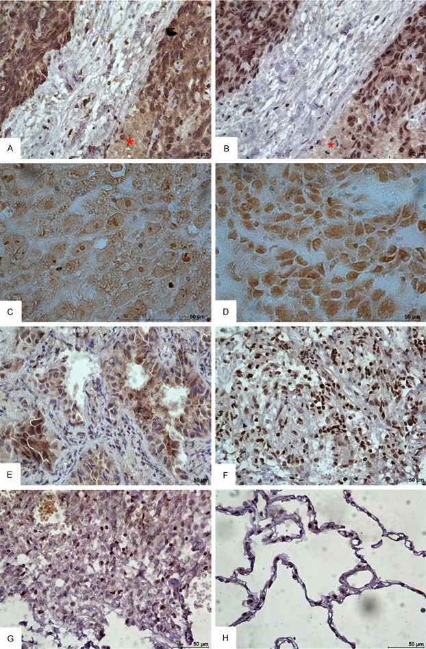
Figure 1.
Representative immunohistochemical staining images for cTnI in human NSCLC tissues (DAB, bar = 50 μm). (A, B) Immunohistochemical staining (with hematoxylin counterstaining) in the lung squamous cell carcinoma tissue from the same patient. (A) Stained with 2F6.6; (B) Stained with 19C7. All showed strong staining. Asterisk showed cellular necrosis area. (C, D) Immunohistochemical staining without hematoxylin counterstaining and with higher magnification. (C) Stained with 2F6.6; (D) Stained with 19C7. (E, F) The tissues from adenocarcinoma and tuberculosis showed positive staining. (E) Adenocarcinoma, (F) Tuberculosis. (G, H) The pulmonary sclerosing hemangioma and non-cancer-bearing lung tissue showed negative staining. (G) Pulmonary sclerosing hemangioma, (H) Non-cancer-bearing lung tissue collected away from tumor area.

