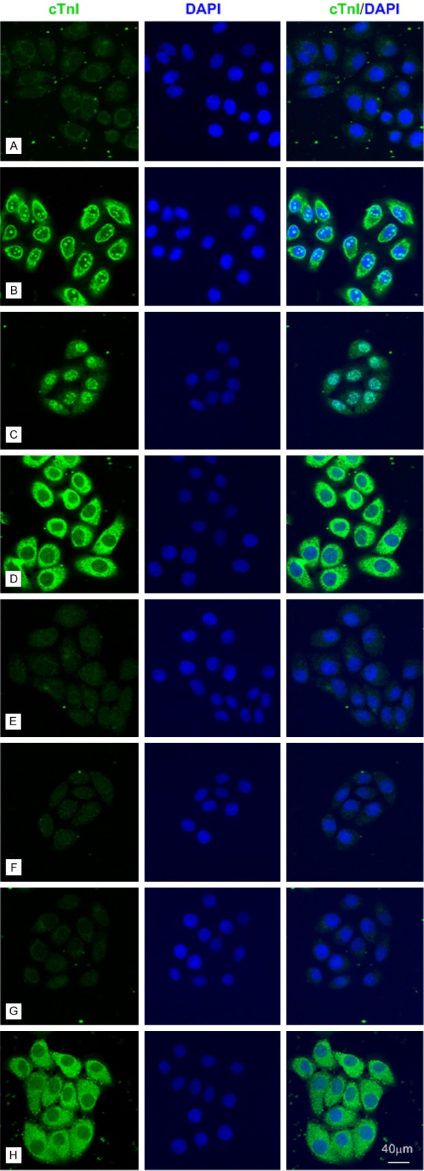
Figure 2.

The anti-cTnI mAbs could stain different parts of SPCA-1 cells by immunofluorescence with an epitope-dependent pattern. Representative confocal images of SPCA-1 cells (bar = 40 μm). A: Negative control. B-H: SPCA-1 cells were stained for cTnI (Fluorescein Avidin DCS, green) by using 2F6.6, 19C7, XY15, 16A11, XY13, 84 and XY10 mAbs, which recognized cTnI epitopes embodied at 13-36, 41-49, 56-80, 86-90, 76-110, 117-126 and 181-210, respectively.
