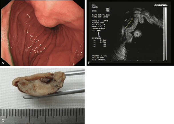
Figure 1.
Representative images of glomus tumor in the stomach. A: Gastroendoscopy shows a round elevated lesion with an overlying normal mucosa in the stomach wall. B: Endoscopic ultrasound image shows a solid mass originating from the superficial layer of the muscular propria. The mass is 0.83 cm × 0.67 cm in size, and a marginal halo is observed. The mass appears as a round, hypoechoic lesion with heterogeneous echogenicity. C: Gross image shows that the tumor is localized in the submucosal area with a clear boundary. The cross-section of the tumor appears gray in color.

