
Figure 3.
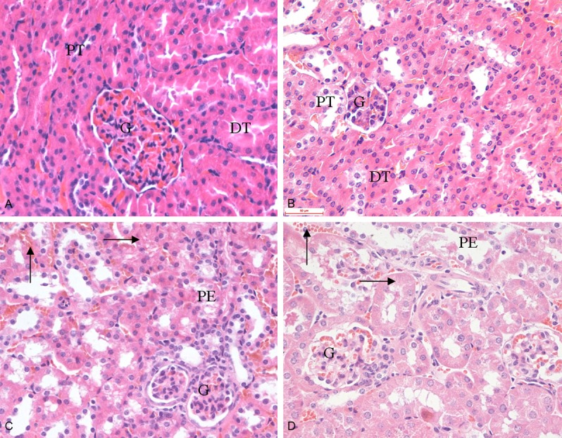
Effect of 1,8-cineole on the microstructures of kidneys in mice after administration for 30 days. A: Group I (0 mg/kg, HE 400×); B: Group II (21.38 mg/kg, HE 400×); C: Group III (64.15 mg/kg, HE 400×); D: Group IV (192.45 mg/kg, HE 400×). A and B: Showed the normal appearance of kidney, glomerulus (G), proximal tubule (PT) and distal tubule (DT). C and D: Showed a dose-related haemorrhagia (↑), granular degeneration (→), renal tubular epithelial cells swelling and separated from basement membrane. The renal tubal lumen containing eosinophilic protein exudation (PE).