
Figure 5.
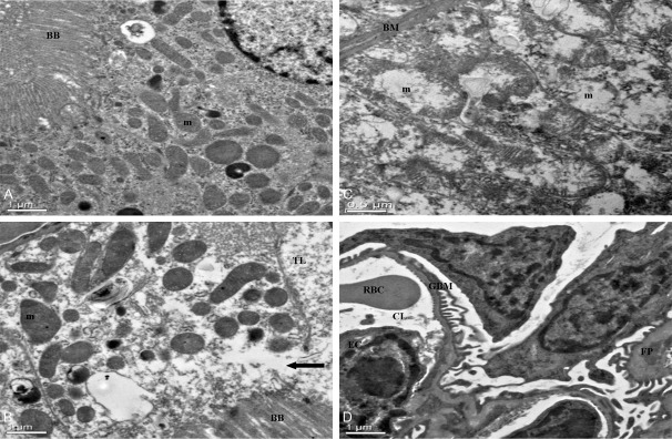
The ultrastructural changes of kidney cells treated with or without 1,8-cineole. A: Normal mice kidney, ×2,550. Brush border (BB), mitochondria (m), Nucleus (N); B: Proximal tubular cell of kidney, 30 days of administrated by 1,8-cineole (192.45 mg/kg). Note swelling and disorganization of mitochondria, and discontinuity of brush border at arrow, with escape of cytoplasm into tubular lumen, ×2,550. Brush border (BB), mitochondrion (m), tubular lumen (TL). C: Distal tubule of kidney, ×6,000. 30 days of administrated by 1,8-cineole (192.45 mg/kg). Note swelling and disorganization of mitochondria (m), the fingerlike projections of the basement membrane (BM) of distal convoluted tubule epithelial cells appeared distorted by the mitochondrial swelling, marked disorganization of the cristae and stippling in the matrix. D: Glomerulus, ×2,550. 30 days of administrated by 1,8-cineole (192.45 mg/kg). The fine structure appears well preserved. Glomerular Basement Membrane (GBM), foot process (FP), endothelial cells (EC), capillary lumen (CL), red blood cell (RBC).