
Figure 1.
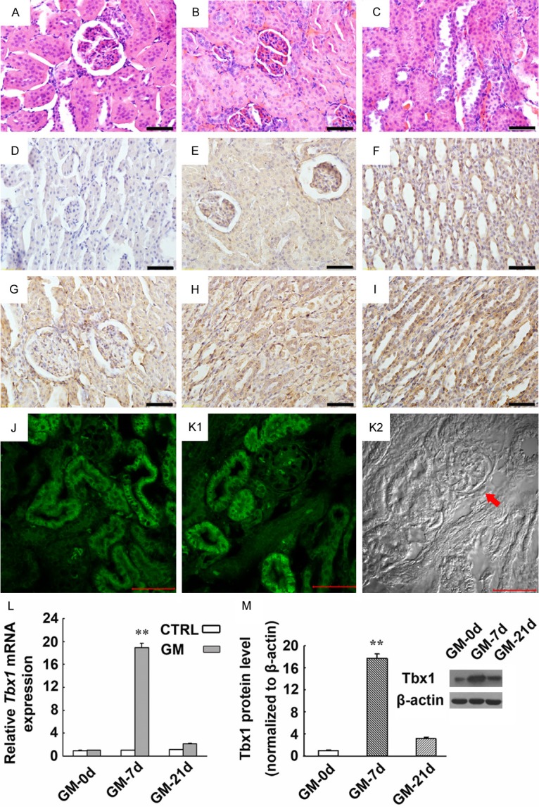
Expression of Tbx1 mRNA and protein in kidney tissues derived from a rat model for AKI induced by GM. (A-C) CTRL rats have shown normal tubular epithelial cells (A). Swollen renal tubular epithelial cells, poorly defined cell boundaries, dissolved nuclei, disappearance of lumen structure (B), and shedding renal tubular epithelial cells were detected in the injured kidneys of the GM-7d group (C) (HE staining). (D-I) Immunohistochemical staining showed the expression and localization of Tbx1 in the kidneys of the GM-7d and CTRL groups. Tbx1 was predominantly expressed in the cytoplasm of renal tubular epithelial cells and collecting duct cells in the cortex and medulla (E-I), which was mild in the kidneys of the CTRL group (E, F), but markedly increased in the GM-7d group (G-I). Magnification: ×400; scale = 50 μm. A negative control is also shown (D). (J-K2) Immunofluorescence staining demonstrated that Tbx1 expression was located predominantly in the cytoplasm of tubular epithelial cells of GM-7d rats (J, K1). The same image in K1 was also captured using DIC microscopy (K2). The arrowhead indicates the glomerulus that is not shown in K1. Magnification: ×400; scale = 50 μm. (L) Tbx1 mRNA expression was significantly up-regulated in the kidneys of the GM-7d group compared with the CTRL, GM-0d and GM-21d groups. (M) Tbx1 protein as detected by Western blotting has increased accordingly compared with the CTRL, GM-0d and GM-21d groups. **, P < 0.01 vs. CTRL, GM-0d and GM-21d groups. No significant changes were found between the GM-0d and GM-21d groups.