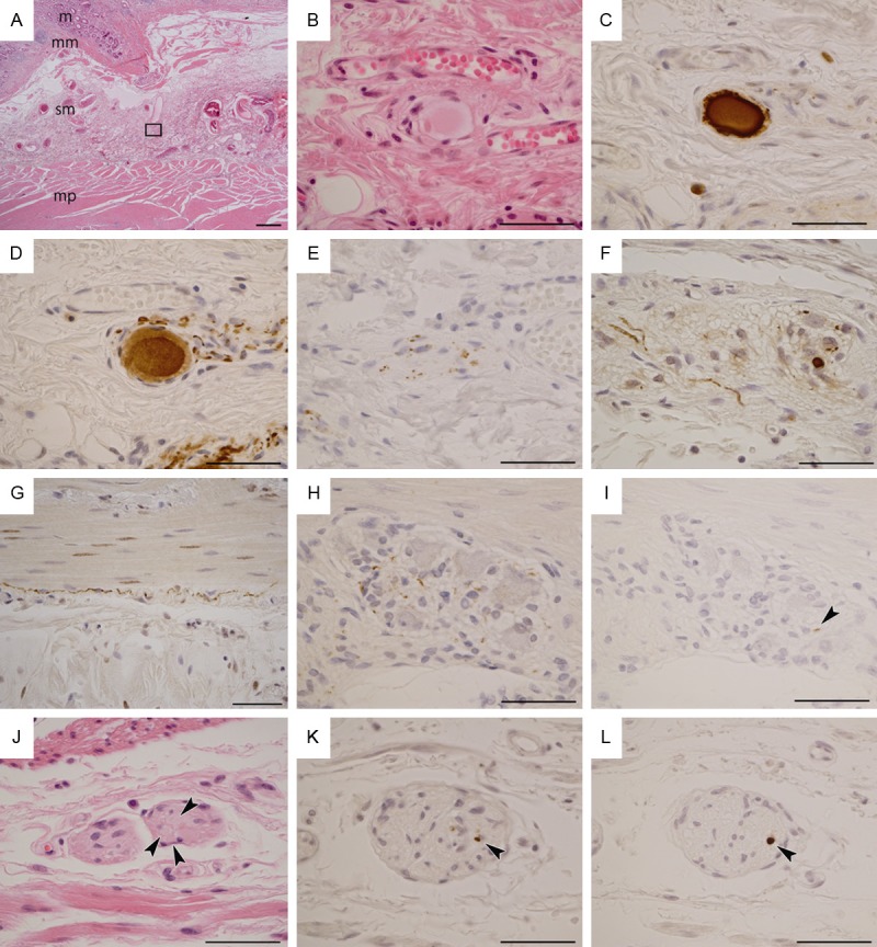
Figure 1.

Photomicrographs of Lewy pathology in surgical specimens of Lewy body disease. See Table 1 for the clinical details of the patients. A-E: Photomicrographs from patient 1. F-I: Photomicrographs from patient 3. J-L: Photomicrographs from patient 6. A: Section of the stomach; mucosa (m), muscularis mucosae (mm), submucosa (sm) and muscularis propria (mp). Rectangle corresponds to panel B, including Meissner’s submucosal nerve plexus. B: An oval-shaped hyaline structure formed in the plexus. C: The hyaline structure shows reactivity against monoclonal phosphorylated α-synuclein antibody (pSyn#64). D: The hyaline structure also shows reactivity against phosphorylated neurofilament (SMI31) antibody. In addition, the structure is apparently located in an SMI31-immunoreactive ganglion cell. E: The plexus shows punctate anti-tyrosine hydroxylase immunoreactivity. F: pSyn#64-immunoreactive neurites and small deposits in Auerbach’s myenteric nerve plexus in the small intestine. G: pSyn#64-immunoreactive long neurites in a subserosal nerve fascicle of the small intestine. H: pSyn#64-immunoreactive small deposits in Auerbach’s myenteric nerve plexus of the small intestine. I: A short and small polyclonal phosphorylated α-synuclein (PSer129)-immunoreactive neurite (arrowhead) in the same plexus as shown in H. J: Arrowheads indicate round hyaline bodies (Lewy bodies) in Auerbach’s myenteric nerve plexus of the surgically removed stomach. K: A small pSyn#64 immunoreactive deposit in Auerbach’s myenteric nerve plexus of the stomach (arrowhead). L: A PSer129 immunoreactive round region in the same plexus as shown in K (arrowhead). A: Scale bar = 500 μm; B-L: Scale bar = 50 μm. A, B, J: Hematoxylin and eosin staining. D: SMI31; E: Tyrosine hydroxylase; C, F, G, H, K: pSyn#64; I, L: PSer129.
