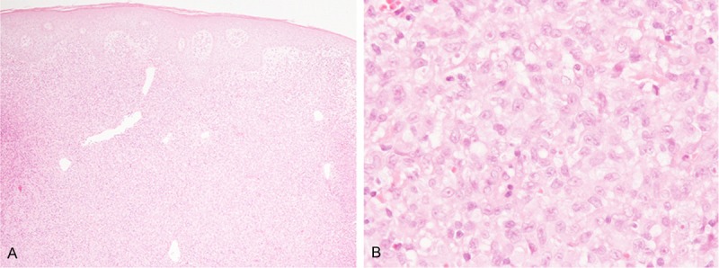
Figure 1.

Histopathological features of the first biopsy specimen from the abdominal wall. A: Diffuse proliferation of atypical lymphocytes in the dermis without epidermotropic infiltration, HE, × 40. B: Proliferation of large-sized lymphocytes with large convoluted nuclei with conspicuous nucleoli and relatively rich eosinophilic cytoplasm, HE, × 400.
