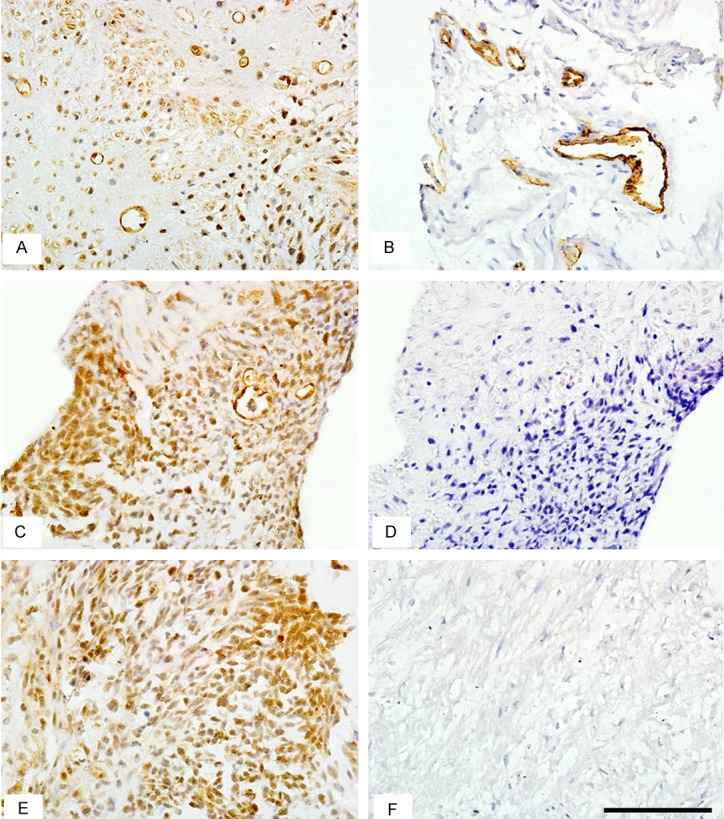
Figure 3.

Immunohistochemical staining of the tumor cells. Immunostaining of the lesion displayed strong reactivity for CD34 (A) and diffuse positivity for CD117 (C) and DOG-1 (E). (B) is a positive control for CD34 staining illustrating the vessels, while (D) and (F) are blank controls for CD117 and DOG-1, respectively. (magnification: × 400; Scale bars = 100 μm).
