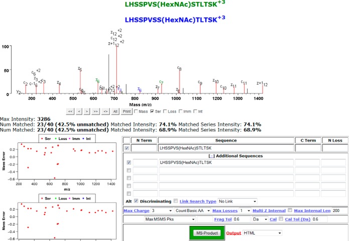
Fig. 2.
Example of MS-Viewer displaying an annotated spectrum for a peptide in which the modification site localization was ambiguous. The peptide has two potential HexNAc modification site localizations shown in different colors; those peak assignments common to both are labeled in black, and those assignments unique to one or the other are labeled in the relevant color.

