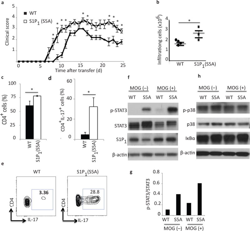
Figure 3. Enhanced STAT3-mediated Th17 polarization in S1P1(S5A) EAE mice.

(a) Mean clinical scores ((±S. E. M.) of Rag1−/− adoptive transfer recipients of encephalitogenic cells from MOG35–55-immunized S1P1(S5A)and WT (C57BL/6J) EAE mice. Donors, n=10, females, 8–9 weeks old; recipients, n=5, females, 5–6 weeks old. (b) Quantification of total CNS infiltrating immune cells, (c) CD4+ and (d, e) CD4+IL-17+ cells from CNS of S1P1(S5A)and WT EAE mice (at peak disease, days 13–15). Immunoblot depicting p-STAT3 (f), p-p38, p38 and IκBα (h) expression in splenocytes of S1Ps1(S5A) and WT EAE mice (day8, post-immunization). (g) Quantification of relative p-STAT3 expression [form (f)]. (b-g) n=3–5 mice/arm. *p<0.05, Mann-Whitney U-test (a) and Student’s t-test (b–d). These experiments were repeated twice (a) and at least 3 times (b–g).
