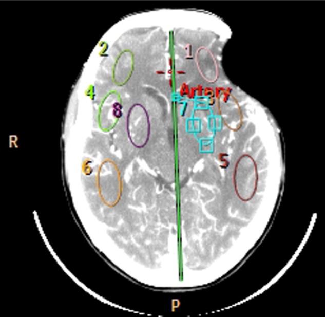
Figure 1.

Computed tomography perfusion with four regions of interest selected in each cerebral hemisphere.
Note: Three superficial regions and one positioned in the basal ganglia.
Abbreviations: R, right; P, posterior.

Computed tomography perfusion with four regions of interest selected in each cerebral hemisphere.
Note: Three superficial regions and one positioned in the basal ganglia.
Abbreviations: R, right; P, posterior.