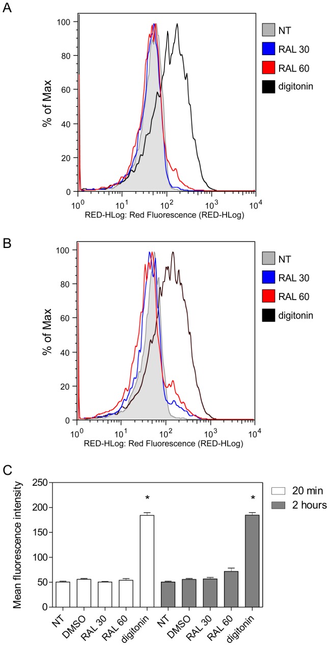
Figure 3. Propidium iodide labeling after raloxifene treatment.

Parasites were incubated at 25°C in M199 medium, left untreated or treated with 30 or 60 µM raloxifene and analysed upon addition of propidium iodide. Parasites treated with 25 µM digitonin were used as a positive control. Untreated parasites and parasites incubated with the highest volume of drug diluent (DMSO 0.6%) were used as negative controls. Fluorescence histograms are representative of three independent experiments with untreated parasites (gray), 30 µM raloxifene (blue), 60 µM raloxifene (red) or 25 µM digitonin (black) for 20 min (A) or 2 hours (B). (C) Values shown are the mean fluorescence intensity ± standard deviation of three independent experiments. (*) indicates significant difference relative to the untreated group (p<0.0001). NT: untreated; RAL: raloxifene.
