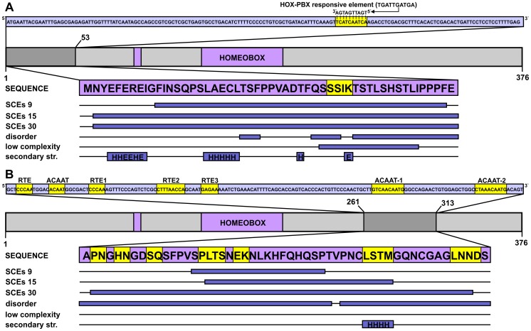
Figure 3. DNA-level secondary functions in coding regions: The case of the HOXA2 gene.
The homeobox protein Hox-A2 is represented by a light grey bar, with its sole domain (homeobox) and antp-type motif colored purple (residue boundaries assigned based on the UniProtKB annotation) and its SCE-overlapping N-terminal region marked by dark grey. The CDS corresponding to this segment is shown above the domain map in a light blue box with the region of multi-functionality (a HOX-PBX responsive element) highlighted in yellow. The corresponding peptide sequence is presented in a purple box with the precise locations of detected SCEs, predicted disordered regions, low sequence complexity segments and secondary structure elements (H – helix, E – extended) represented as dark blue bars below the protein sequence. B) The enhancer-rich region corresponding to residues 261–313 of the same Hox protein is presented in a similar fashion as in panel A.

