
Figure 4. HAT inhibition affects the normal development of S. mansoni eggs.
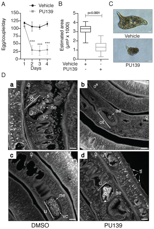
Ten adult worm pairs were cultivated with 20 µM PU139 or vehicle up to four days, and the number of eggs was counted on a daily basis (A). The estimated areas (length and width) of these same eggs were measured (B). Student's t-test was applied, with ***p<0.001. The morphology of the eggs was analyzed under optical microscopy. Scale bars: 10 µm (C). Adult worm pairs under the same treatment were fixed and stained with hydrochloric carmine for confocal laser scanning microscopy (CLSM) analysis. ne: normal egg; ae: abnormal egg; ot: ootype; vc: vitelline cells; u: uterus. The arrowhead in “ae” points to a fissure. tt: tegument tubercles. Scale bars: 20 µm (D).