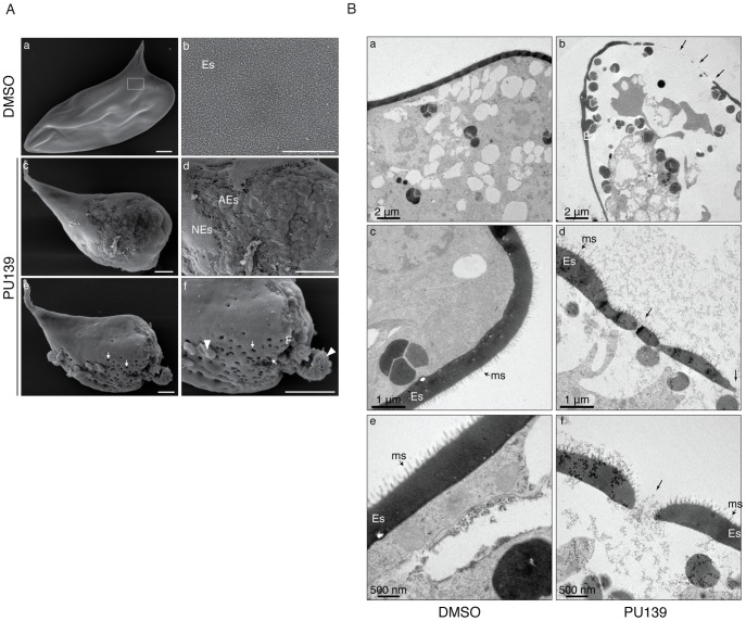
Figure 5. HAT inhibition compromises eggshell integrity.
(A) Scanning electron microscopy of eggs laid by worm pairs cultivated for two days with vehicle (panels a and b) or 20 µM PU139 (panels c, d, e and f). A normal S. mansoni egg is shown (panel a) with the typical smooth coat of microspines on the eggshell (Es, panel b) seen at higher magnification (boxed area in panel a). PU139 treatment severely affected eggshell formation and integrity (panel c). Gross structural defects on the eggshell can be visualized at higher magnification (panel d; NEs: normal eggshell, AEs: abnormal eggshell). A remarkable phenotypic defect observed in eggs laid by PU139-treated parasites was the presence of holes in the eggshell (arrows in panel e). At a higher magnification, it can be clearly observed that, besides the holes (panel e and f, arrows), a large fissure in the eggshell was formed (panel f, F), leading to leakage of egg contents (panel f, arrowheads). Scale bars 10 µm. (B). Transmission electron microscopy of eggs laid by worm pairs cultivated for two days with vehicle (panels a, c and e) or 20 µM PU139 (panels b, d, and f). Normal eggs (panels a, d and e) show a thick and continuous eggshell (Es). The characteristic microspines (ms) of S. mansoni eggs are indicated by the arrows. The eggshells of the eggs laid by PU139-treated females (panels b, d and f) revealed remarkable differences in their structure, when compared with the control eggs, showing much thinner and discontinuous (arrows) eggshells. The holes in the eggshells were confirmed by TEM.

