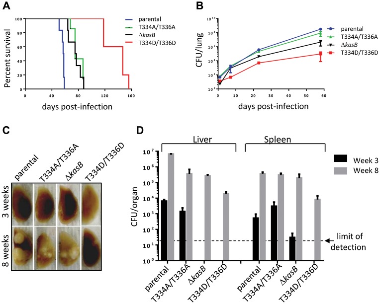
Figure 5. Infection of immunocompromised SCID mice with M. tuberculosis kasB isogenic mutants. (A) Survival curves in infected SCID mice.
Low-dose aerosol infection of SCID mice (as in B) was performed with the following Mtb strains: parental, KasB_T334A/T336A, KasB_T334D/T336D and ΔkasB. (B) Growth of Mtb kasB strains in the lungs of SCID mice. At 1, 7, 21 and 56 days post-infection, one lung from each infected SCID mouse was harvested, homogenized and serial dilutions were plated on Middlebrook 7H10 supplemented with 10% OADC and 0.2% glycerol. (C) Pathology of lungs from infected SCID mice. One lung from each infected SCID mouse was harvested and fixed in 10% paraformaldehyde for a month prior to photography. (D) CFU plots in the liver and spleen three weeks (black bars) and eight weeks (grey bars) post-infection.

