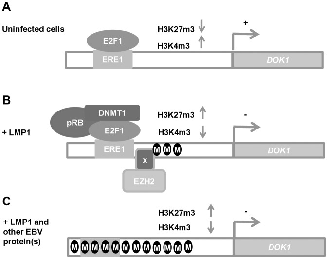
Figure 7. Schematic model of DOK1 gene regulation in EBV-infected cells.
(A) In uninfected cells, DOK1 expression is activated via the recruitment of the active form of the E2F1 transcription factor to its response element located at (−498/−486) on the DOK1 promoter. (B) In cells expressing the oncoprotein LMP1, DOK1 is down-regulated through the recruitment of the inhibitory complexes E2F1/pRB/DNMT1 and EZH2 to its promoter region. These complexes lead to the induction of partial DNA methylation and the increase of H3K27 trimethylation levels, respectively. (C) In EBV-infected cells, DOK1 is repressed through heavy DNA methylation of its promoter region and the increase in H3K27 trimethylation level. These events likely induce conformational changes in the chromatin, which become less permissive to E2F1 transcription factor recruitment.

